What is a Neck Dissection?
The purpose of a neck dissection is to remove the lymph nodes in the neck. The surgery is almost always done for certain types of head and neck cancer. (Head and neck cancer is often described as throat cancer, but it can also include cancer of the mouth, the tongue, the thyroid gland, the saliva glands, etc.)
Cancer in this region can spread through the lymphatic system to adjacent lymph nodes. If this happens, a neck dissection can be done to remove the suspicious lymph nodes. There approximately 600 lymph nodes in the body, and 200 of this are located in the neck. A neck dissection is useful not only to remove the cancer, but also so that the nodes can be examined by a pathologist. If the microscopic examination by the pathologist shows extensive spread of cancer, additional treatment such as radiation therapy may be recommended.
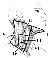
 This image shows the various regions of the neck where the lymph nodes are located. The neck is typically divided into zones (zones I through VI are shown on this diagram). Research has shown that tumors tend to follow certain trends in the manner in which the spread. For example, tumors of the mouth tend to spread first to the upper neck zone ( e.g. zones I, II, and III). Tumors lower in the neck, for example laryngeal cancer, tend to spread to lower zones (zones III or IV).
How a neck dissection is done
A neck dissection is done through an incision in the neck. There are many important anatomical structures, such as nerves and blood vessels, that run through the neck, and these structures are carefully identified and preserved in the course of the operation. If the original (also called primary) tumor is going to be treated with surgery, this tumor resection is usually done at the same time as the neck dissection. For example, if the primary site of the tumor is the larynx (voicebox), a part or all of the larynx may be removed at the same time as the neck dissection.
Important neck structures
There are three other important structures in the neck that are closely involved in a neck dissection. These are the internal jugular vein, the spinal accessory nerve, and the sternocleidomastoid muscle. The internal jugular vein is a major blood vessel through which blood returns from the head to the heart. The spinal accessory nerve controls movement of some of the major shoulder muscles. The sternocleidomastoid muscle is a large muscle in the neck.
During a neck dissection the surgeon will try to save these structures. However, if the tumor invades into one or all of these structures, one or all of them will have to be removed.
Types of neck dissections
Neck dissections are classified by the zones from which the lymph nodes are removed and whether or not the three structures described above are preserved. If all the nodes are removed (zones I through V) and the three structures (internal jugular vein, accessory nerve and sternocleidomastoid muscle) are removed it is called a radical neck dissection (radical is a misleading term; it just means that a complete neck dissection has been done). A radical neck dissection would be done if the tumor spread to the neck is quite extensive. If the nodes from zones I through V are removed and one of these three structures is preserved, it is called a modified radical neck dissection. And if the operation does not involve all five zones, it is called a selective neck dissection.
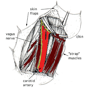
 The image to the left shows the appearance of the neck after a radical neck dissection. The sternocleidomastoid muscle, the internal jugular vein, and the spinal accessory nerve have been removed. The surgeon has saved the vagus nerve (which supplies the muscles in the throat and the larynx) and the hypoglossal nerve (which supplies the muscles that move the tongue). Also preserved is the carotid artery, which is extremely important for providing oxygen to the brain. The strap muscles are strap-shaped muscles that help raise and lower structures in the neck. Note that skin "flaps" have been elevated, which allows very good exposure to all the structures in the neck. Exposure is very important in surgery. The important structures to be saved must first be identified, and exposure is key to finding and protecting these nerves and blood vessels.
What to expect after surgery
Recovery time from a neck dissection alone can be short. If a patient has only had a neck dissection, the total hospital stay may only be several days. However, if the surgery is done along with resection of the primary tumor site, this can take longer. Considering only the neck dissection, there usually will be drains placed under the skin at the time of surgery to collect any serous fluid or blood that accumulates. These drains will be removed after a couple of days. Once they are out, and if the incision is healing well, the patient can usually go home.
It is not unusual to develop some numbness in the neck skin and ear after a neck dissection. Much of the numbness goes away after several months, but some may be permanent. The nerve supplying sensation to the ear often must be cut during the surgery and some of this sensation never returns. If you live in a cold climate, you will have to be careful since a numb ear can develop frostbite without first feeling cold or painful. The remaining numbness can be bothersome but seldom causes any major problems.
Effect of removing neck structures
One or more neck structures (internal jugular vein, spinal accessory nerve, or sternocleidomastoid muscle) may need to be removed in a neck dissection. Removal of one jugular vein usually causes minimal or no problems. There are many other veins in the neck and the blood can flow back through them. There may be some temporary swelling, but this usually decreases after a couple weeks. If a neck dissection is being done on both sides of the neck, the surgeon will try to save at least one jugular vein. Both veins can be removed at the same time, but the consequences of the swelling can be quite severe.
Removal of the spinal accessory nerve limits the upward movement of the shoulder. In particular, it will be more difficult to move the arm from the horizontal position to directly overhead. There also may be some shoulder droop on the side of surgery and there can be some mild pain due inflammation at the shoulder joint. If the spinal nerve is removed, post-operative physical therapy will be crucial to maintain good shoulder function.
Removal of one sternocleidomastoid muscle (SCM) generally causes no problems, however, the neck may look a little sunken due to its removal. The muscle also provides some coverage of the carotid artery, so this will be reduced if the muscle is resected. This can cause a problem if there is a post-operative infection, but otherwise it is not that important. If both sternocleidomastoid muscles are removed, one's strength in flexing the head forward will be reduced.
Other possible complications of a neck dissection
There are many important structures running through the neck and any one of these can be injured during a neck dissection. Nerves to the lower lip and the tongue are in the operative field, and injury may lead to reduced movement of the tongue. If this weakness occurs, it usually is temporary. Another nerve supplies some of the muscles in the lower lip and these can become weak after the surgery.
As with any surgery there are risks of bleeding or infection after surgery. If the bleeding or infection is severe, it may be necessary to go back to the operating room to treat the problem. The operation is done under a general anesthetic. If there are pre-existing lung or heart problems this can increase the risk of a pneumonia or heart attack.
It is important to remember the risk of a major complication after a neck dissection is relatively small and almost always are far outweighed by the benefits of the operation.
